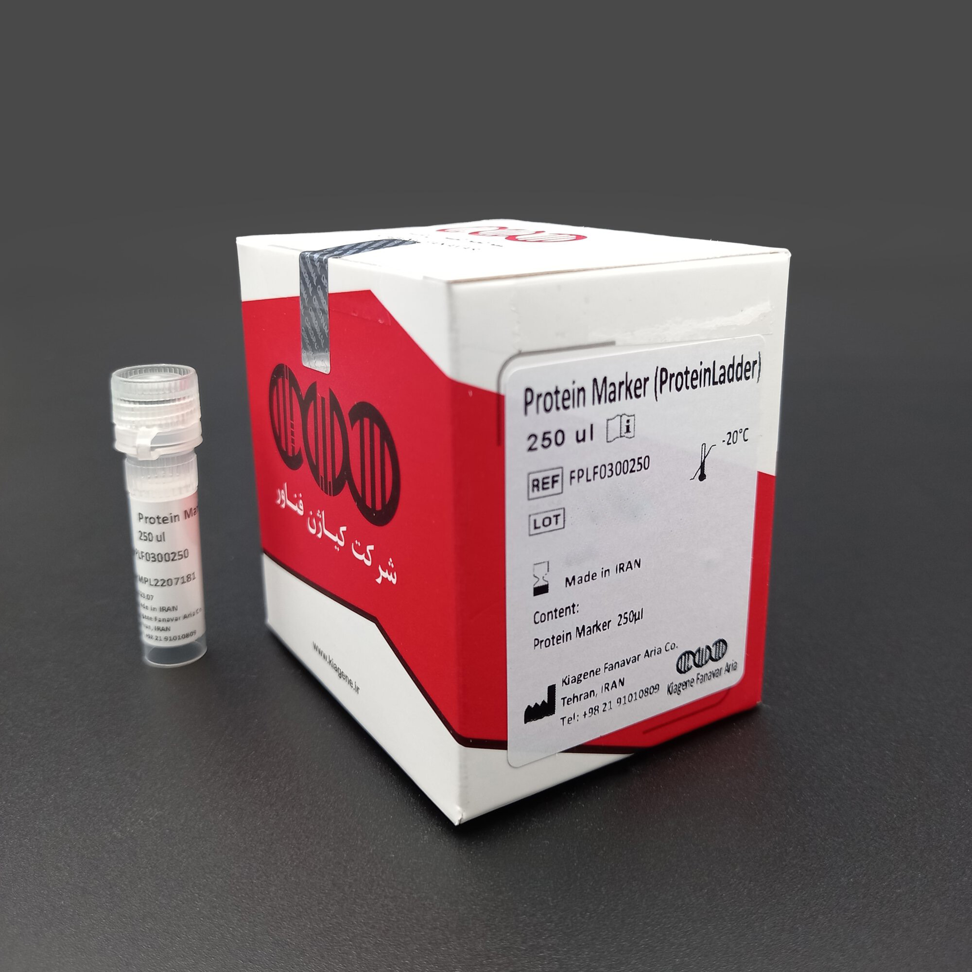
نشانگر- لدر- پروتئین|Protein Marker-ladder

نشانگر- لدر- پروتئین|Protein Marker-ladder


حجم موجود
0.1mL
- تاریخ انقضا حداقل یک سال
- نگهداری 20- درجه سانتی گراد
15,000,000 ریال







توضیحات محصول
نشانگر- لدر- پروتئین|Protein Marker-ladder
Protein Marker-ladder
Cat. No:
FPLF030.0100
Contents:

Kit storage:
 This kit should be stored at -20 °C.
If properly stored, all kit components are stable until the expiration date printed on the label.
Additional Equipment and Reagent required
- Electrophoresis apparatus (gel tank, comb, glass plates, and power supply)
- Pre-cast or handcast polyacrylamide gels (SDS-PAGE)
- Protein loading buffer
- Reducing agent (β-mercaptoethanol)
- Electrophoresis running buffer (Tris-Glycerol-SDS- β-mercaptoethanol – Bromophenol blue)
- Protein staining solution (e.g., Coomassie Brilliant Blue R-250 or G-250, Silver stain, or compatible fluorescent stain)
- Destaining solution (Methanol/Acetic acid or as per staining kit instructions)
- Gel imaging system or scanner
- Microcentrifuge tubes and pipettes with tips
- Heating block or water bath (95-100°C) for sample denaturation
- Deionized or distilled water
Application
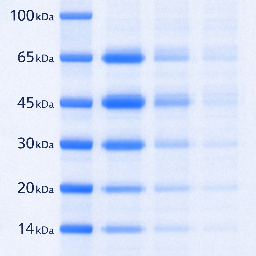
The Protein Marker-ladder is designed for use as a molecular weight standard in SDS-Polyacrylamide gel electrophoresis (SDS-PAGE) and Western blotting applications utilizing a post-staining detection method.This ladder consists of six distinct, highly purified recombinant proteins with molecular weights of 14, 20, 30, 45, 65, and 100 kDa. Upon electrophoresis and subsequent staining, it produces sharp, well-defined bands, allowing for accurate estimation of the molecular mass of unknown sample proteins.
Kiagene protein Marker – protein ladder
The specific protein sources for each band are as follows:
14 kDa Band: Lysozyme (from chicken egg white)
20 kDa Band: Beta-lactoglobulin (or Trypsin inhibitor, from bovine milk/soybean)
30 kDa Band: Carbonic Anhydrase (from bovine erythrocytes)
45 kDa Band: Ovalbumin (from chicken egg white)
65 kDa Band: Albumin (Bovine Serum Albumin – BSA)
100 kDa Band: Phosphorylase B (from rabbit muscle)
This range is particularly suitable for monitoring protein separation during electrophoresis, verifying transfer efficiency onto membranes, and estimating the size of target proteins in complex biological samples.
Handling Requirements and Safety Information
Upon receipt, store the Protein Marker-ladder immediately at -20°C.
Under proper storage conditions, the product is stable until the expiration date stated on the label.
For routine use, aliquot the marker into smaller volumes to avoid repeated freeze-thaw cycles, which may degrade the protein bands. It is recommended to store working aliquots at 4°C for short-term use (up to 1-2 weeks) and return the stock to -20°C.
If precipitation is observed, briefly warm the marker to 37°C-40°C and mix gently before use
This product is intended for research use only (RUO) and is not for diagnostic or therapeutic use in humans or animals.
Always wear appropriate personal protective equipment (PPE), including laboratory coats, safety goggles, and gloves, when handling the product.
Avoid contact with skin, eyes, and clothing. In case of contact, flush immediately with copious amounts of water and seek medical attention if irritation persists.
Do not ingest or inhale. If swallowed, seek medical advice immediately and show the container or label.
Dispose of all waste in accordance with local, state, and federal environmental regulations.
Protocol
Protocol for Use (SDS-PAGE with Post-Staining)
This protocol is optimized for the Protein Marker-ladder using a post-staining detection method.
A. Preparation of Sample Loading Buffer (2X Sample Loading Buffer Formulation)
- 125 M Tris-HCl (pH 6.8)
- 4% SDS (w/v)
- 20% Glycerol (v/v)
- 2% 2-Mercaptoethanol (2-ME) (v/v)
- 01% Bromophenol Blue (w/v)
Preparation Instructions:
- Prepare 0.125 M Tris-HCl buffer and adjust to pH 6.8.
- Dissolve SDS, Glycerol, and Bromophenol Blue in the buffer.
- Add 2-Mercaptoethanol just before use, as it is volatile and degrades over time.
- Store aliquots at -20°C for long-term stability.
B. Sample and Marker Preparation
- Thawing: Thaw the Protein Marker-ladder and your protein samples at room temperature or on ice.
- Mix your protein sample with the 2X Sample Loading Buffer in a 1:1 ratio
- Heat the sample mixtures at 95-100°C for 5-10 minutes.
- Briefly centrifuge to collect condensate.
- Marker Preparation: Vortex the marker gently for 2-3 seconds. Briefly centrifuge the tube to collect the contents at the bottom.
C. Gel Electrophoresis
- Gel Setup: Assemble the electrophoresis apparatus and insert a pre-cast or handcast polyacrylamide gel (SDS-PAGE). Fill the chambers with the appropriate running buffer (e.g., Tris-Glycine-SDS).
Loading:
- Carefully load 5-10 µL of the Protein Marker-ladder into the first and/or last well.
- Load your prepared protein samples (mixed with loading buffer) into the remaining wells.
Running Conditions: Run the gel at a constant voltage (e.g., 100-200V) until the dye front (Bromophenol Blue) reaches the bottom of the gel.
D. Post-Electrophoresis Staining (Post-Stain)
Gel Removal: After electrophoresis, carefully transfer the gel into a clean container filled with deionized water.
Fixing (Recommended): Incubate the gel in a fixing solution (e.g., 40% Methanol, 10% Acetic acid) for 30-60 minutes with gentle shaking.
Staining:
For Coomassie Blue Staining:
- Pour off the fixing solution and add enough Coomassie staining solution (e.g., 0.1% Coomassie Brilliant Blue R-250 in 40% Methanol, 10% Acetic acid) to cover the gel.
- Incubate for 30-60 minutes at room temperature with gentle agitation.
For Silver Staining or Fluorescent Stains: Follow the manufacturer’s instructions.
Destaining:
- Remove the staining solution and incubate the gel in destaining solution (e.g., 40% Methanol, 10% Acetic acid) with gentle shaking. Change the solution 2-4 times until the background is clear and the marker bands are visible.
E. Visualization and Analysis
- Imaging: Capture the gel image using a gel documentation system.
- Analysis: Locate the six distinct bands (14, 20, 30, 45, 65, and 100 kDa) to estimate the molecular weight of your sample proteins.
بررسی تخصصی
سایر ویژگیها
حجم به میکرولیتر را انتخاب کنید





احسان –
عالی و درجه یک
inhiple –
In an in vivo tumor xenograft model, mice are implanted with tumors
inhiple –
Incomeppc KbMoRmCKkwpXdChr 5 20 2022 In addition to the normal complications of cysts, the presence of cysts in pregnancy may cause obstructed labor
inhiple –
3Max DelbrГјck Centrum fГјr Molekulare Medizin MDC and NeuroCure, Berlin, Germany
ignitonew –
Biomedis Tamoxifen indications